How Advanced Craniofacial Surgery Helped Transform the Life of a Child With Crouzon Syndrome
A Story That Extends Beyond Appearance
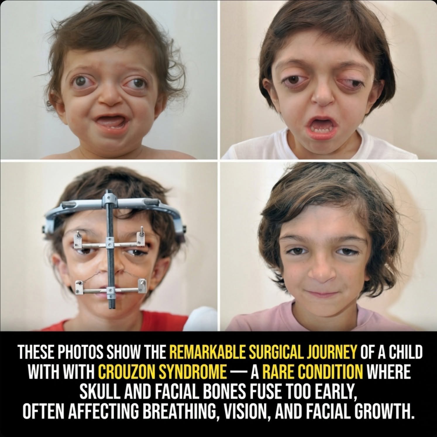
Medical advancements are often explained through technical language, research papers, and clinical discussions. Yet sometimes a series of photographs can communicate the impact of a treatment more clearly than pages of scientific terminology.
Images documenting the transformation of a child living with Crouzon syndrome have captured widespread attention because of the remarkable physical changes visible over time.
While many people naturally focus on the differences in facial appearance, the most significant achievement of the treatment is not cosmetic.
The true success lies in restoring critical functions that affect breathing, vision, sleep, and overall quality of life.
Beneath the visible transformation is a highly specialized surgical process designed to address serious structural challenges caused by a rare genetic condition.
The procedure represents a combination of medical expertise, biological healing, and advanced surgical planning.
For children affected by severe forms of Crouzon syndrome, these interventions can dramatically improve daily life and long-term health outcomes.
Understanding Crouzon Syndrome
Crouzon syndrome is a rare genetic disorder that affects the development of the skull and facial bones.
The condition is characterized by craniosynostosis, a process in which certain bones of the skull fuse earlier than they should during childhood development.
Under normal circumstances, the skull remains flexible while the brain continues to grow.
These growth patterns allow the head and facial structures to expand gradually throughout early childhood.
In children with Crouzon syndrome, some of these bones fuse prematurely.
As a result, normal growth becomes restricted.
The condition creates a mismatch between the body’s developmental needs and the physical limitations imposed by the fused bones.
Although the brain continues to grow, the surrounding skeletal structure may not provide enough room to accommodate that growth naturally.
This imbalance can affect multiple areas of the face and skull.
The Challenges Created by Premature Bone Fusion
The effects of Crouzon syndrome extend far beyond appearance.
Because parts of the skull and midface stop developing normally, several important structures can become compressed or underdeveloped.
The facial skeleton often lacks sufficient forward growth.
This condition, known as midface deficiency, can significantly alter the shape and function of the face.
One of the most noticeable consequences involves the eye sockets.
When the surrounding bones remain shallow, the eyes may appear unusually prominent.
This prominence is not simply a cosmetic concern.
Shallow orbital cavities can reduce the natural protection provided to the eyes.
The condition may increase vulnerability to irritation, injury, and other complications.
At the same time, the underdevelopment of facial bones can affect the nasal passages and upper airway.
These spaces may become narrowed, making it difficult for a child to breathe normally.
Tasks that most people take for granted, such as breathing comfortably during sleep, can become significant challenges.
How Airway Restriction Affects Daily Life
Restricted airways represent one of the most serious medical concerns associated with severe midface deficiency.
When the upper airway becomes too narrow, breathing may require constant effort.
Children can experience ongoing discomfort and fatigue as their bodies work harder to obtain adequate airflow.
Nighttime often presents additional difficulties.
Many children with severe airway narrowing develop obstructive sleep apnea.
This condition occurs when airflow becomes partially or completely blocked during sleep.
Repeated interruptions in breathing can reduce sleep quality and place additional stress on the body.
Over time, inadequate oxygen levels and disrupted sleep may affect overall health and development.
For families, these challenges can become a daily concern.
Simple activities such as sleeping, exercising, and maintaining normal energy levels may be affected.
Addressing these functional problems is one of the primary goals of surgical treatment.
The Role of Modern Craniofacial Surgery
Advances in craniofacial medicine have created new possibilities for treating severe facial skeletal abnormalities.
Among the most important procedures used in cases of significant midface deficiency is a technique known as a Le Fort III distraction.
This approach combines precise surgical planning with the body’s natural ability to generate new bone.
Rather than forcing immediate and dramatic repositioning of facial structures, the procedure allows gradual correction over time.
The method is carefully designed to support healing while producing meaningful anatomical improvements.
It represents a sophisticated blend of surgical science and biological regeneration.
The procedure requires careful coordination among specialized medical teams.
Every stage is planned with the goal of improving both structure and function.
The First Step: Separating the Midface
The process begins with a carefully controlled surgical procedure.
Surgeons create precise cuts that separate the central facial skeleton from the surrounding skull.
This section includes important structures such as the cheekbones, upper jaw, and nasal bridge.
The separation is performed with extraordinary accuracy.
Every movement must be planned to preserve vital tissues while creating the conditions necessary for future correction.
The objective is not merely to alter appearance.
The goal is to reposition an entire region of the facial skeleton so that critical anatomical functions can improve.
This stage establishes the foundation for the next phase of treatment.
Using Controlled Movement to Create Change
After the facial bones are separated, surgeons attach a specialized external device known as a distractor.
This frame serves as the mechanism that gradually guides the bones into a new position.
Instead of moving the facial skeleton all at once, adjustments occur slowly over several weeks.
The device is modified by extremely small amounts each day.
These tiny movements are often measured in fractions of a millimeter.
Although the changes may appear minimal on a daily basis, they accumulate steadily over time.
The gradual approach allows the body to adapt while minimizing stress on surrounding tissues.
It also creates an environment that encourages natural bone formation.
This controlled movement is one of the most remarkable aspects of the procedure.
How the Body Creates New Bone
As the distractor slowly moves the facial bones forward, a small gap develops between the separated sections.
Rather than leaving an empty space, the body responds through its natural healing process.
New bone tissue begins to form within the gap.
This biological response allows the skeletal structure to lengthen gradually while maintaining stability.
The process is known as distraction osteogenesis.
It relies on the body’s own regenerative capabilities rather than artificial replacement materials.
As healing continues, healthy new bone develops and matures.
The result is a lasting structural change supported by the patient’s own tissue.
This combination of engineering principles and biological healing has transformed the treatment of complex craniofacial conditions.
Expanding the Space Around the Eyes
One of the major objectives of the procedure is increasing the volume of the orbital cavities.
When the midface moves forward, the eye sockets also expand.
This additional space allows the eyes to rest in a more protected position.
The improvement helps address one of the hallmark features associated with severe Crouzon syndrome.
Beyond appearance, enhanced protection for the eyes can reduce risks linked to exposure and inadequate support.
The change represents an important functional benefit.
By improving the surrounding anatomy, surgeons help create safer conditions for long-term eye health.
This outcome illustrates how structural corrections can directly influence essential biological functions.
Opening the Airway
Perhaps the most important effect of advancing the midface is the expansion of the upper airway.
As the facial skeleton moves forward, the space available for airflow increases.
This improvement can significantly reduce airway obstruction.
Children who previously struggled with restricted breathing may experience substantial relief.
The benefits often become especially apparent during sleep.
Improved airflow can lessen the impact of obstructive sleep apnea and reduce interruptions in breathing.
Families frequently notice improvements in rest, energy levels, and overall comfort.
The ability to breathe more freely is one of the most meaningful outcomes of treatment.
For many patients, this functional improvement becomes the defining success of the entire surgical journey.
Measuring Success Beyond Photographs
Before-and-after images can be powerful because they reveal visible evidence of change.
However, the most important results often cannot be captured in a single photograph.
The true measure of success is found in everyday experiences that become easier after treatment.
A child who can breathe comfortably through the night experiences benefits that extend far beyond appearance.
Peaceful sleep, improved oxygen flow, and reduced physical strain can influence many aspects of health and development.
These gains represent the deeper purpose behind complex craniofacial procedures.
While visual transformation may draw public attention, functional improvement remains the central objective.
Every stage of treatment is designed with that goal in mind.
The Intersection of Compassion and Innovation
The treatment of severe craniofacial conditions demonstrates what can be achieved when medical expertise and human compassion work together.
Surgeons, nurses, specialists, and families collaborate through a lengthy process that often requires patience and dedication.
Each step reflects a commitment to improving a child’s quality of life.
The science involved is highly advanced, but the purpose is deeply human.
Helping a child breathe more easily, sleep more peacefully, and live more comfortably remains the ultimate objective.
Modern craniofacial surgery has expanded the possibilities available to patients facing complex developmental conditions.
Techniques such as Le Fort III distraction illustrate how biological healing can be guided through carefully planned medical engineering.
The results stand as a powerful example of how innovation can address challenges that once seemed impossible to overcome.
A Remarkable Achievement in Modern Medicine
The transformation seen in children treated for severe Crouzon syndrome represents far more than a change in facial appearance.
It reflects the correction of structural problems that can affect breathing, vision, sleep, and overall wellbeing.
Through the combination of precise surgery, gradual skeletal movement, and natural bone regeneration, physicians can create meaningful improvements in both function and quality of life.
The process highlights the extraordinary relationship between anatomy and survival.
Facial structures do much more than shape appearance. They play a critical role in protecting the eyes, maintaining open airways, and supporting essential bodily functions.
When those structures develop abnormally, the consequences can be profound.
When modern medicine successfully restores them, the impact can be life-changing.
The journey of a child treated for Crouzon syndrome serves as a powerful reminder of how far medical science has advanced and how those advancements continue to transform lives every day.